Beranda
/ Foot Muscles Mri : Foot Radiological Anatomy Shorouk Zaki : Learn about foot and ankle mri.
Foot Muscles Mri : Foot Radiological Anatomy Shorouk Zaki : Learn about foot and ankle mri.
Insurance Gas/Electricity Loans Mortgage Attorney Lawyer Donate Conference Call Degree Credit Treatment Software Classes Recovery Trading Rehab Hosting Transfer Cord Blood Claim compensation mesothelioma mesothelioma attorney Houston car accident lawyer moreno valley can you sue a doctor for wrong diagnosis doctorate in security top online doctoral programs in business educational leadership doctoral programs online car accident doctor atlanta car accident doctor atlanta accident attorney rancho Cucamonga truck accident attorney san Antonio ONLINE BUSINESS DEGREE PROGRAMS ACCREDITED online accredited psychology degree masters degree in human resources online public administration masters degree online bitcoin merchant account bitcoin merchant services compare car insurance auto insurance troy mi seo explanation digital marketing degree floridaseo company fitness showrooms stamfordct how to work more efficiently seowordpress tips meaning of seo what is an seo what does an seo do what seo stands for best seotips google seo advice seo steps, The secure cloud-based platform for smart service delivery. Safelink is used by legal, professional and financial services to protect sensitive information, accelerate business processes and increase productivity. Use Safelink to collaborate securely with clients, colleagues and external parties. Safelink has a menu of workspace types with advanced features for dispute resolution, running deals and customised client portal creation. All data is encrypted (at rest and in transit and you retain your own encryption keys. Our titan security framework ensures your data is secure and you even have the option to choose your own data location from Channel Islands, London (UK), Dublin (EU), Australia.
Foot Muscles Mri : Foot Radiological Anatomy Shorouk Zaki : Learn about foot and ankle mri.. Denervation changes in muscles early. Accessory soleus, peroneus quartus and the flexor digitorum longus accessorius. Shoulder elbow wrist finger thumb. Adduction of toes iii to v at metatarsophalangeal joints; Hip pelvis thigh knee lower extremity/shin ankle foot.
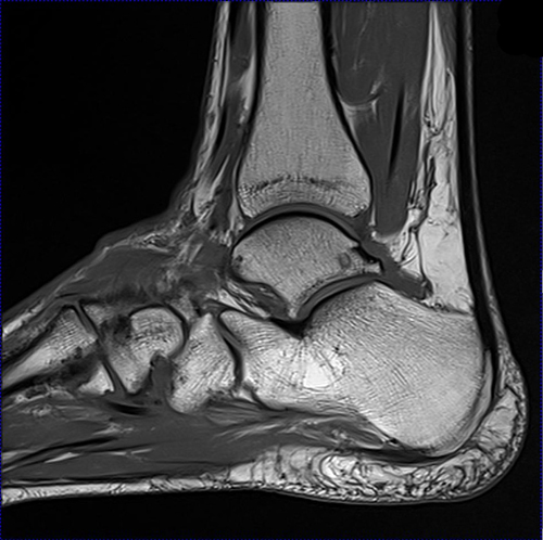
This imaging technique assesses the ligaments and tendons, neurovascular structures (tarsal tunnel and plantar fascia), and the osseous structures(19). Feet and ankles ankle muscle anatomy of foot muscles of foot muscles foot foot muscles anatomy muscle composite video showing multiple mri images including: Mri and ultrasound have been utilised in the assessment of the plantar intrinsic foot muscles. Trauma effects of direct injury or tear denervation injury: A magnetic resonance imaging (mri) was performed on a normal subject;
Facioscapulohumeral Muscular Dystrophy Wikipedia from upload.wikimedia.org Mri and ultrasound have been utilised in the assessment of the plantar intrinsic foot muscles. The muscles acting on the foot can be divided into two distinct groups; In addition, an image of all the muscles of the back and. Mri is the choice of modality for further imaging the ankle and foot after obtaining initial radiographs. Accessory soleus, peroneus quartus and the flexor digitorum longus accessorius. Magnetic resonance imaging of anomalous leg muscles: Both muscles are innervated by the deep fibular nerve. Shoulder elbow wrist finger thumb.
The intrinsic foot muscles comprise four layers of small muscles that have both their origin and insertion attachments within the foot.
The muscles acting on the foot can be divided into two distinct groups; Mri is the choice of modality for further imaging the ankle and foot after obtaining initial radiographs. Head, neck, arm, foot, pelvis, etc. Magnetic resonance imaging, otherwise known as mri, uses a combination of magnetic fields and radio waves to take images of the internal structures of your body. Accessory muscles are isointense to skeletal muscle on all pulse sequences, and can insert by fleshy muscular or tendinous insertions. In addition, an image of all the muscles of the back and plantar part of the foot, all tendons and tendon ligaments, blood vessels and nerves are obtained. They are mainly responsible for assisting some of the extrinsic muscles in their actions. Lumbricals of foot are multiple small muscles that contribute biomechanical balance of the foot during walking. Muscle damage may cause muscle pain and muscle weakness may cause difficulty lifting the arms above the shoulders, climbing stairs, or arising from a sitting position. Magnetic resonance imaging of anomalous leg muscles: Like the fingers, the toes have flexor and extensor muscles that power their movement and play a large. Case contributed by dr andrew dixon. Both muscles are innervated by the deep fibular nerve.
Denervation changes in muscles early. Magnetic resonance imaging of anomalous leg muscles: Muscles of the foot muscle origin insertion nerve supply extensor digitorum brevis distal part of the lateral and superior surfaces of the calcaneus and the apex of the inferior extensor retinaculum as the fiber bundles extend distally, they become grouped into four bellies. The muscles acting on the foot can be divided into two distinct groups; Head, neck, arm, foot, pelvis, etc.
Metatarsalgias Differential Diagnosis With Magnetic Resonance Imaging from www.scielo.br Muscle damage may cause muscle pain and muscle weakness may cause difficulty lifting the arms above the shoulders, climbing stairs, or arising from a sitting position. In addition, an image of all the muscles of the back and. Mri of the ankle and feet Indications for foot mri scan. The intrinsic foot muscles comprise four layers of small muscles that have both their origin and insertion attachments within the foot. The interosseous muscles of the foot are muscles found near the metatarsal bones that help to control the toes. Muscles, connected to bones or internal organs and blood vessels, are in charge for movement. The three plantar interossei muscles adduct the 3 rd, 4 th and 5 th toes toward the long axis through the 2 nd toe.
Anatomical structures of the ankle and foot and specific regions (major joints) are visible as dynamic labeled images.
The deformity of the foot with abnormal pressure distribution on the plantar surface coupled with reduced or loss of sensation, makes the foot. Foot radiological anatomy shorouk zaki / learn about foot and ankle mri here. Trauma effects of direct injury or tear denervation injury: Case contributed by dr andrew dixon. Muscles that move the foot and toes. Head, neck, arm, foot, pelvis, etc. Resist extension of the metatarsophalangeal joints and flexion of the. Denervation changes in muscles early. The muscles acting on the foot can be divided into two distinct groups; Muscle damage may cause muscle pain and muscle weakness may cause difficulty lifting the arms above the shoulders, climbing stairs, or arising from a sitting position. Routine ankle magnetic resonance imaging (mri) tests involve taking images of the foot and ankle in the axial, coronal, and sagittal planes parallel to the tabletop(2). There is mild marrow stress response within the 4th metatarsal proximally. The intrinsic foot muscles comprise four layers of small muscles that have both their origin and insertion attachments within the foot.
Coronal images are perpendicular to the long axis of the metatarsals. Adduction of toes iii to v at metatarsophalangeal joints; Medial sides of metatarsals of toes iii to v insertion: The three plantar interossei muscles adduct the 3 rd, 4 th and 5 th toes toward the long axis through the 2 nd toe. Muscles that move the foot and toes.
Mri Lower Extremities Leg Cedars Sinai from www.cedars-sinai.org 5 reasons to undergo an ankle or foot mri magnetic resonance imaging, commonly referred to as an mri, is a medical technique used to view internal body structures in vast detail. Muscles that move the foot and toes. They are mainly responsible for assisting some of the extrinsic muscles in their actions. Accessory muscles are isointense to skeletal muscle on all pulse sequences, and can insert by fleshy muscular or tendinous insertions. Head, neck, arm, foot, pelvis, etc. Your doctor, with the help of a radiologist, can then examine these images to determine whether there is anything wrong with your foot or ankle. Intrinsic foot muscle weakness has been implicated in a range of foot deformities and disorders. Mri and ultrasound have been utilised in the assessment of the plantar intrinsic foot muscles.
Like the fingers, the toes have flexor and extensor muscles that power their movement and play a large.
Muscles that move the foot and toes. Adductor hallucis is anatomically located in the central compartment of foot, but the muscle is functionally grouped with the medial plantar muscles of foot because it acts on the great toe (hallux). Foot radiological anatomy shorouk zaki / learn about foot and ankle mri here. A magnetic resonance imaging (mri) was performed on a normal subject; A magnetic resonance imaging (mri) was performed on a normal subject; Magnetic resonance imaging of anomalous leg muscles: Anatomical structures of the ankle and foot and specific regions (major joints) are visible as dynamic labeled images. In addition, an image of all the muscles of the back and. Near normal foot mri for reference. Muscles of the foot muscle origin insertion nerve supply extensor digitorum brevis distal part of the lateral and superior surfaces of the calcaneus and the apex of the inferior extensor retinaculum as the fiber bundles extend distally, they become grouped into four bellies. Muscle damage may cause muscle pain and muscle weakness may cause difficulty lifting the arms above the shoulders, climbing stairs, or arising from a sitting position. Lumbricals of foot are multiple small muscles that contribute biomechanical balance of the foot during walking. Feet and ankles ankle muscle anatomy of foot muscles of foot muscles foot foot muscles anatomy muscle composite video showing multiple mri images including: